


 Foundational characteristics of cancer include proliferation, angiogenesis, migration, evasion of apoptosis, and cellular immortality. Find key markers for these cellular processes and antibodies to detect them.
 The SUMOplot™ Analysis Program predicts and scores sumoylation sites in your protein. SUMOylation is a post-translational modification involved in various cellular processes, such as nuclear-cytosolic transport, transcriptional regulation, apoptosis, protein stability, response to stress, and progression through the cell cycle.
 The Autophagy Receptor Motif Plotter predicts and scores autophagy receptor binding sites in your protein. Identifying proteins connected to this pathway is critical to understanding the role of autophagy in physiological as well as pathological processes such as development, differentiation, neurodegenerative diseases, stress, infection, and cancer.
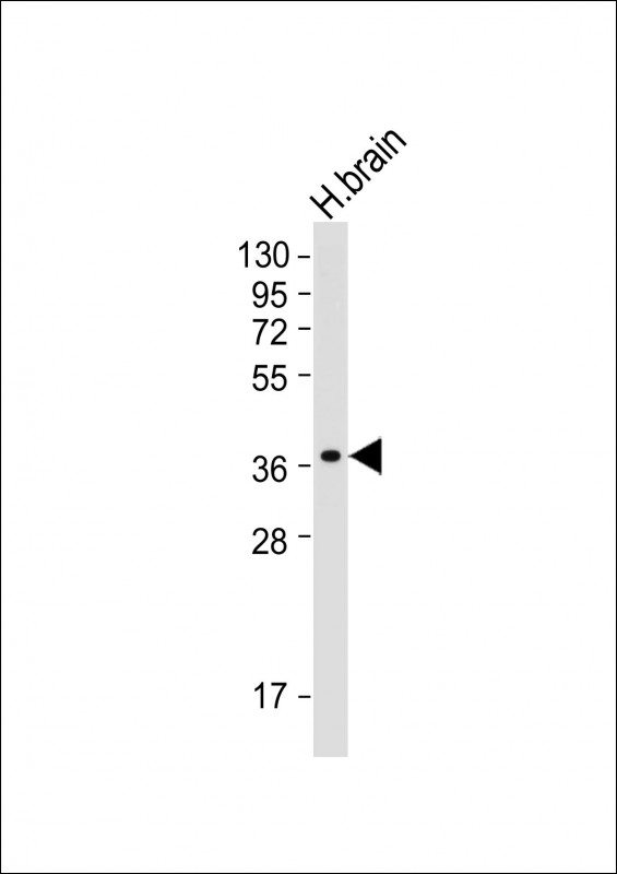
GDE1 Antibody
Purified Mouse Monoclonal Antibody (Mab)
- SPECIFICATION
- CITATIONS
- PROTOCOLS
- BACKGROUND

Application
| WB, E |
|---|---|
| Primary Accession | Q9NZC3 |
| Reactivity | Human |
| Predicted | Human |
| Host | Mouse |
| Clonality | monoclonal |
| Isotype | IgG1,κ |
| Clone/Animal Names | 2021CT823.61.7 |
| Calculated MW | 37718 Da |
| Gene ID | 51573 |
|---|---|
| Other Names | Glycerophosphodiester phosphodiesterase 1, 3.1.4.44, Membrane-interacting protein of RGS16, RGS16-interacting membrane protein, GDE1, MIR16 |
| Target/Specificity | This GDE1 antibody is generated from a mouse immunized with a recombinant protein from the human region of human GDE1. |
| Dilution | WB~~1:2000 E~~Use at an assay dependent concentration. |
| Format | Purified monoclonal antibody supplied in PBS with 0.09% (W/V) sodium azide. This antibody is purified through a protein G column, followed by dialysis against PBS. |
| Storage | Maintain refrigerated at 2-8°C for up to 2 weeks. For long term storage store at -20°C in small aliquots to prevent freeze-thaw cycles. |
| Precautions | GDE1 Antibody is for research use only and not for use in diagnostic or therapeutic procedures. |
| Name | GDE1 (HGNC:29644) |
|---|---|
| Function | Hydrolyzes the phosphodiester bond of glycerophosphodiesters such as glycerophosphoinositol (GroPIns) and glycerophosphoethanolamine (GroPEth), to yield a glycerol phosphate and an alcohol (By similarity). Hydrolyzes glycerophospho-N-acylethanolamines to N- acylethanolamines in the brain and participates in bioactive N- acylethanolamine biosynthesis such as anandamide (an endocannabinoid), N-palmitoylethanolamine (an anti-inflammatory), and N- oleoylethanolamine (an anorexic). In addition, has a lysophospholipase D activity by hydrolyzing N-acyl-lysoplasmenylethanolamine (N-acyl- lysoPlsEt) to N-acylethanolamine. However lysophospholipase D activity is lower than glycerophosphodiester phosphodiesterase activity (By similarity). Has little or no activity towards glycerophosphocholine (By similarity). |
| Cellular Location | Cell membrane {ECO:0000250|UniProtKB:Q9JL55}; Multi-pass membrane protein. Cytoplasmic vesicle membrane {ECO:0000250|UniProtKB:Q9JL55}; Multi-pass membrane protein. Note=Perinuclear vesicles and cell membrane {ECO:0000250|UniProtKB:Q9JL55} |
| Tissue Location | Widely expressed.. |

Thousands of laboratories across the world have published research that depended on the performance of antibodies from Abcepta to advance their research. Check out links to articles that cite our products in major peer-reviewed journals, organized by research category.
info@abcepta.com, and receive a free "I Love Antibodies" mug.

Provided below are standard protocols that you may find useful for product applications.
Background
Has glycerophosphoinositol phosphodiesterase activity. Has little or no activity towards glycerophosphocholine. GDE1 activity can be modulated by G-protein signaling pathways (By similarity).
References
Zheng B.,et al.Proc. Natl. Acad. Sci. U.S.A. 97:3999-4004(2000).
Duennebier F.F.,et al.Submitted (NOV-2003) to the EMBL/GenBank/DDBJ databases.
Loftus B.J.,et al.Genomics 60:295-308(1999).
Bachmann A.S.,et al.Gene 371:144-153(2006).
If you have used an Abcepta product and would like to share how it has performed, please click on the "Submit Review" button and provide the requested information. Our staff will examine and post your review and contact you if needed.
If you have any additional inquiries please email technical services at tech@abcepta.com.
Ordering Information
Other Products
Shipping Information
